From analog to digital
- All dissection slides are digitized
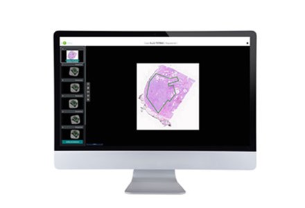
- ROI annotation transfer algorithm
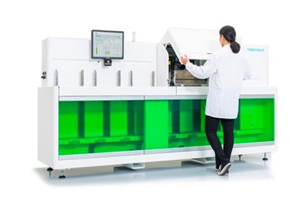
- Robotized dissection
 From manual to automated
- Automatic annotation transfer
- Automatic dissection
- Slides in - Sample tubes out
 From black box to documented
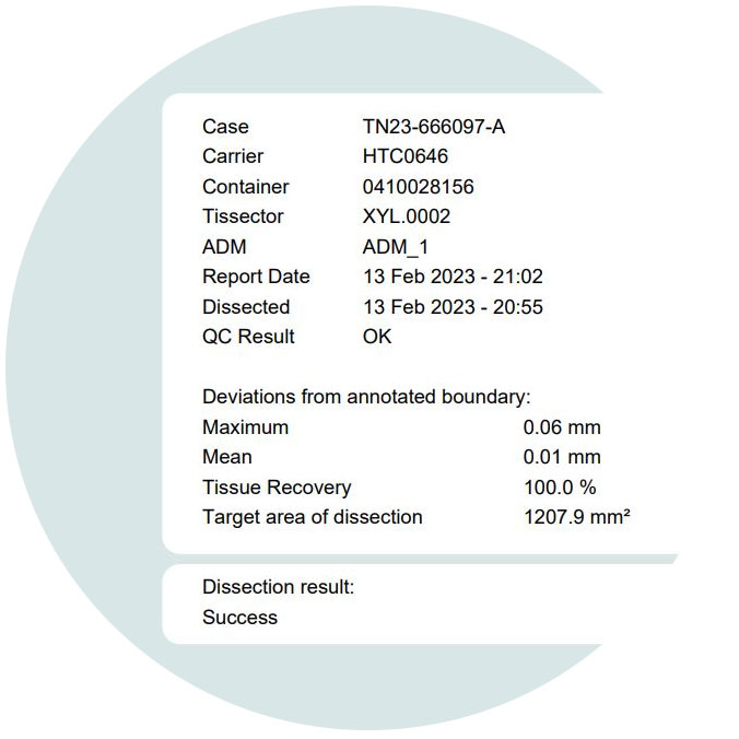
- QC report including pre- and post-dissection images
- Fully integrated with LIMS and IMS
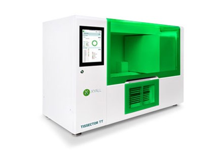
Tissector workflow
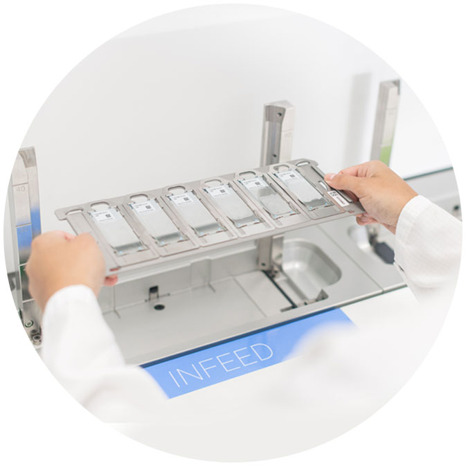
Slide loading
All barcoded slides are placed on a carrier that is inserted in the instrument.
Slide scanning
All dissection slides are digitized and images are sent to the Annotation Suite.
Region of Interest annotation
All images are made available per case to the pathologist in the Annotation Suite, for ROI selection and marking, supported by easy to use annotation tools.
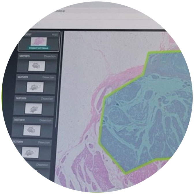
Annotation transfer
The marked ROIs are automatically transferred to the dissection slide images by using the latest image registration technologies. The result can be reviewed by the pathologist and adapted if necessary.
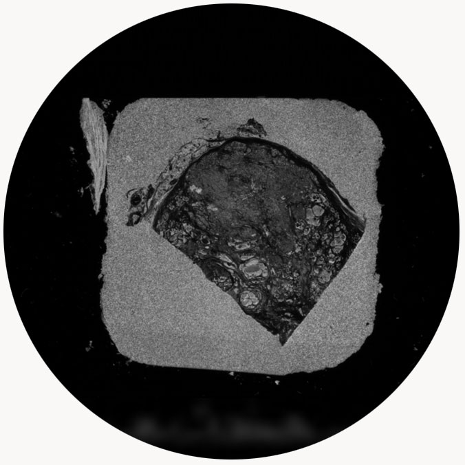
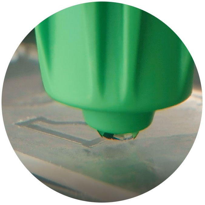
Tissue dissection
The Tissector instrument scrapes the tissue automatically from the dissection slides with high precision using a consumable scraping head. For each case a new scraping head is picked up automatically.
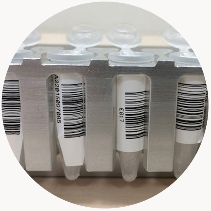
Process output
After dissection the collected tissue is transferred automatically into designated sample tubes ready for the molecular work up. No liquids are used in the process.
Quality Control
Images are taken before and after scraping to check and document the accuracy of the dissection. A QC report is generated including images and uploaded to the LIMS.